Kidney stones are one of the most common urinary tract disorders, affecting between 4 and 8% of the Australian population at any given time (Kidney Health Australia 2020).
What are Kidney Stones?
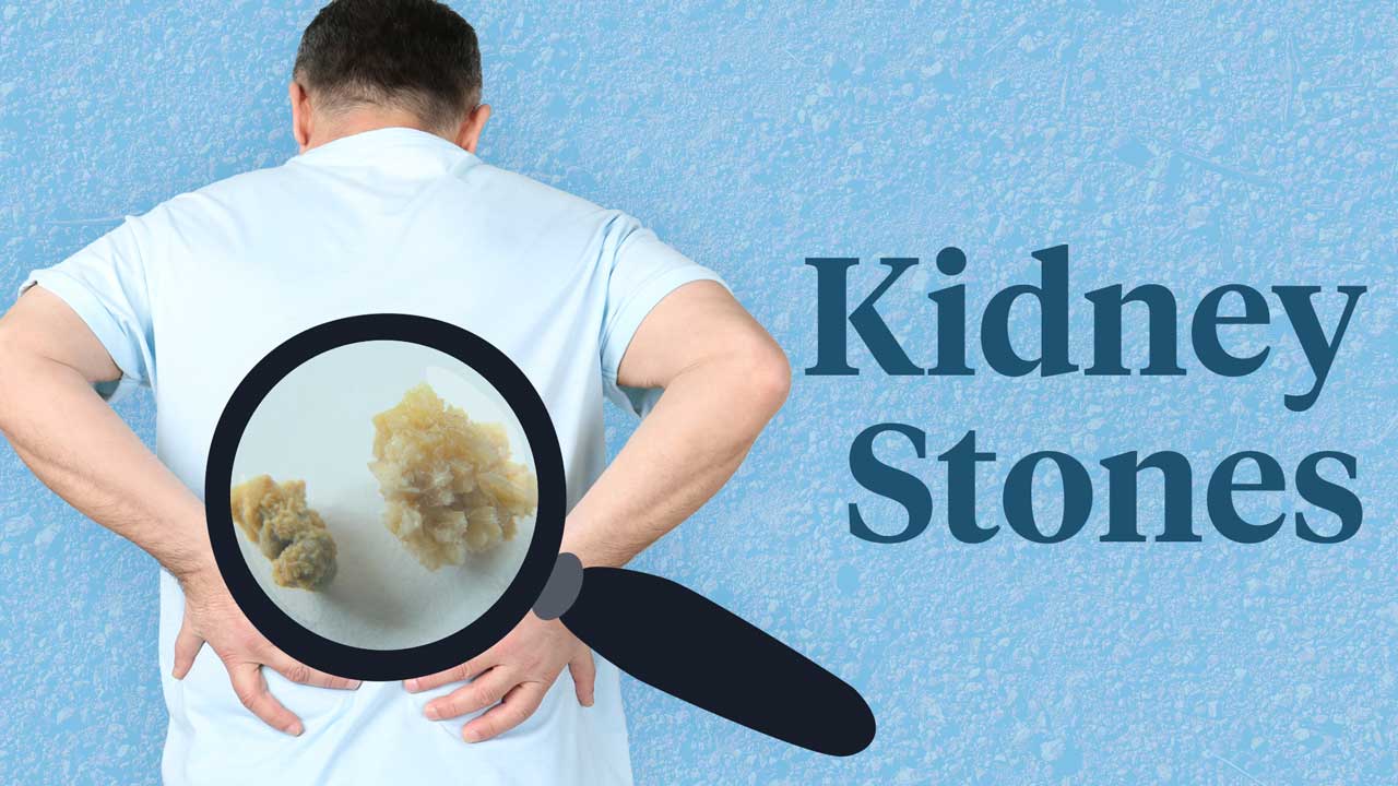
Kidney stones, also known as renal calculi, are crystallised masses formed by salts in the urine (Better Health Channel 2022).
They can range in size from as small as a grain of sand to as large as a golf ball (Healthdirect 2023).
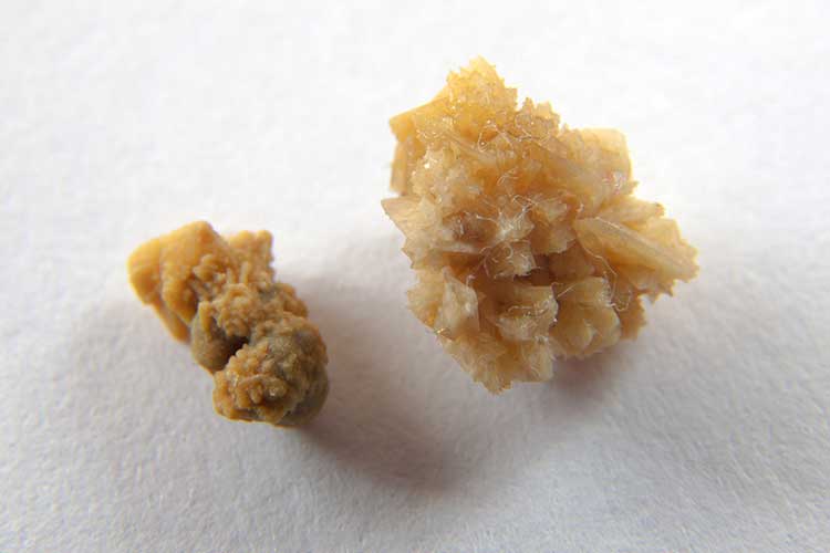
Kidney stones are typically yellow or brown in colour and can be smooth or jagged (Queensland Health 2017).
About 1 in 10 men and 1 in 35 women will develop a kidney stone during their lifetime (Better Health Channel 2022).
What Causes Kidney Stones?
Kidney stones occur when waste products in the urine clump together into hard crystals (Kidney Health Australia 2020).
This can occur for a variety of reasons, including:
- Poor hydration, leading to low urine volume
- Urinary retention
- Dietary factors (e.g. high intake of sodium or oxalate)
- Urinary tract infection
- Systemic acidosis
- Genetic factors
- Taking certain medicines, including some medicines used to treat HIV (atazanavir and indinavir)
- Hypercalciuria (high levels of calcium in the urine)
- Hyperoxaluria (high levels of oxalate in the urine)
- Hyperuricosuria (high levels of uric acid in the urine)
- Hypocitraturia (low levels of citrate in the urine).
(Leslie et al. 2024)

Types of Kidney Stones
There are four types of kidney stones:
- Calcium stones are formed when calcium combines with oxalate or phosphate in the urine. They are the most common type of kidney stone and may be associated with poor calcium and fluid intake or conditions such as hyperparathyroidism, renal calcium leak, hyperoxaluria, hypomagnesemia and hypocitraturia.
- Uric acid stones are typically caused by a high intake of purine-containing foods such as meat, fish and legumes, which results in monosodium urate production. They are also associated with acidic urine (pH of under 5), gout and cancer. Uric acid stones often run in families.
- Struvite stones, which comprise magnesium and ammonia, form after an upper urinary tract infection caused by Gram-negative, urease-producing bacteria, such as Pseudomonas, Proteus and Klebsiella.
- Cystine stones are rare and hereditary. They are caused by a metabolic defect.
(Better Health Channel 2022; Leslie et al. 2024; NKF 2024, 2025a)
Symptoms of Kidney Stones
Kidney stones typically become painful if they pass into the ureter (the tubes connecting the kidneys and bladder) and cause irritation or blockage. While this can cause severe pain, most kidney stones can pass through the body without causing damage to the internal structures (Mayo Clinic 2025; NKF 2025a).
Despite this, many people with kidney stones experience no symptoms (Better Health Channel 2022).
As a general rule, the larger the kidney stone, the more likely it is to cause symptoms (NKF 2025a).
Possible symptoms include:
- Renal colic - severe pain on either side of the lower back, which may move to the front of the body or towards the groin and can fluctuate in intensity
- Nausea or vomiting
- Haematuria (blood in urine)
- Urinary urgency
- Cloudy or foul-smelling urine
- Small, gravel-like stones visible in the urine.
(NKF 2025a; Better Health Channel 2022; Mayo Clinic 2025)

Signs of systemic infection include fever, chills and sweating. This can be potentially life-threatening (Leslie et al. 2024).
Complications of Kidney Stones
Kidney stones have the potential to cause long-term kidney damage. They may also increase the risk of urinary or kidney infection, which could potentially lead to sepsis (Better Health Channel 2022; NHS 2022).
Those who have had one kidney stone are also at increased risk of developing another one in the future. The likelihood of experiencing a second kidney stone within five to seven years is 50% (NKF 2025a).
Diagnosing Kidney Stones
Diagnosis will involve:
- Taking the patient’s history
- Physical examination
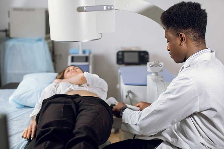
- Imaging (CT scan or kidney-ureter-bladder x-ray) to determine the size and position of the stone
- Urine and blood tests to determine the patient’s kidney health.
(NKF 2025a)
Treating Kidney Stones
In most cases, kidney stones can be treated non-surgically. Most stones will pass on their own within three to six weeks, with about 90% of stones smaller than 5 mm passing with the aid of medical expulsion therapy (Better Health Channel 2022; Leslie et al. 2024).
While small stones can usually be left to pass on their own without any issues, the patient may require analgesia for pain relief and, potentially, intravenous hydration and antiemetic medicines (Leslie et al. 2024).
The patient may also be advised to increase their fluid intake (NKF 2025a).
If the stone is too large or is obstructing urine flow, or the patient is displaying signs of infection, surgical intervention will be required (NKF 2025a). There are several methods of surgical removal, including:
- Extracorporeal shock-wave lithotripsy (ESWL) for stones smaller than 2 cm, which is a non-invasive technique that uses ultrasound waves to break the stone into pieces small enough to pass on their own
- Percutaneous nephrolithotomy/nephrolithotripsy for stones larger than 2 cm, which involves making an incision in the back and removing the stone using specialised instruments. Nephrolithotomy involves removing the stone via a small tube threaded into the incision, while nephrolithotripsy involves breaking the stone into smaller pieces using high-frequency sound waves and then suctioning the fragments through the incision
- Using an endoscope to retrieve the stone or break it into smaller pieces
- Surgical removal of the stone if no other methods are appropriate.
(Better Health Channel 2022; NKF 2025a, b)

The stone should be collected and analysed after it has been passed to determine its cause. The patient may also be advised to collect urine for the next 24 hours so that it can be tested for calcium and uric acid levels. The patient may also undergo blood tests for calcium, phosphorus and uric acid. This will enable a treatment plan to be prescribed that will hopefully prevent the development of future kidney stones (NKF 2025a; Leslie et al. 2024).
When to Escalate Care
Urgent intervention is required in the following situations:
- Pyelonephritis - urinary obstruction combined with urinary tract infection, fever or sepsis
- Nausea or pain that cannot be adequately controlled in an outpatient setting
- An obstructing kidney stone in the patient’s only kidney
- Simultaneous obstruction of both kidneys
- An obstructing kidney stone combined with rising levels of creatinine.
(Leslie et al. 2024)
Preventing Kidney Stones
The risk of kidney stones can be reduced by:
- Reviewing medicines being taken to determine if any of them might be contributing to the risk of developing a stone
- Treating urinary tract infections quickly
- Limiting intake of animal proteins
- Avoiding dehydration and drinking enough fluids to keep a urine volume at or above two litres per day
- Avoiding excessive coffee and tea intake
- Reducing salt intake
- Avoiding excessive intake of drinks containing phosphoric acid (e.g. cola and beer).
(Better Health Channel 2022; Healthdirect 2023)
Test Your Knowledge
Question 1 of 3
What is the most common type of kidney stone?
Topics
References
- Better Health Channel 2022, Kidney Stones, Victoria State Government, viewed 8 May 2025, https://www.betterhealth.vic.gov.au/health/conditionsandtreatments/kidney-stones
- Healthdirect 2023, Kidney Stones, Australian Government, viewed 8 May 2025, https://www.healthdirect.gov.au/kidney-stones
- Kidney Health Australia 2020, Kidney Stones, Kidney Health Australia, viewed 8 May 2025, https://kidney.org.au/your-kidneys/what-is-kidney-disease/types-of-kidney-disease/kidney-stones
- Leslie, SW, Sajjad, H & Murphy, PB 2024, ‘Renal Calculi’, StatPearls, viewed 8 May 2025, https://www.ncbi.nlm.nih.gov/books/NBK442014/
- Mayo Clinic 2025, Kidney Stones, Mayo Clinic, viewed 9 May 2025, https://www.mayoclinic.org/diseases-conditions/kidney-stones/symptoms-causes/syc-20355755
- National Health Service 2022, Kidney Stones, NHS, viewed 9 May 2025, https://www.nhs.uk/conditions/kidney-stones/
- National Kidney Foundation 2024, Uric Acid Stones, NKF, viewed 9 May 2025, https://www.kidney.org/atoz/content/uric-acid-stone
- National Kidney Foundation 2025, Kidney Stones, NKF, viewed 9 May 2025, https://www.kidney.org/atoz/content/kidneystones
- National Kidney Foundation 2025b, Percutaneous Nephrolithotomy/Nephrolithotripsy, NKF, viewed 9 May 2025, https://www.kidney.org/atoz/content/kidneystones_PNN
- Queensland Government 2017, Kidney Stones, Queensland Government, viewed 8 May 2025, http://conditions.health.qld.gov.au/HealthCondition/condition/3/134/431/kidney-stones
 New
New 
