 New
New 

Body temperature is a vital sign used to assess the body’s ability to create and expel heat (Healthwise 2022a).
When measured together with other vital signs, body temperature can help paint an overall picture of a patient’s health and assess circulatory, respiratory, neural, and endocrine functioning (Physiopedia 2022).
Vital signs are highly useful as they are a simple and cost-effective way of gathering information about a patient’s essential physiological functions (Physiopedia 2022).
Measuring a patient’s temperature is specifically useful for identifying possible fever, hyperthermia, or hypothermia (Lapum et al. 2018).
Normal Body Temperature
The normal body temperature range is 36.5°C to 37.5°C (Lapum et al. 2018).
Note: This may vary depending on your organisation's hospital protocol.
However, a person’s temperature can fluctuate based on a number of factors, including:
- Age, as older adults have more difficulty conserving heat. Babies and young children have less effective heat control mechanisms and therefore have a wider temperature range.
- The menstrual cycle, as body temperature is affected by hormones.
- Time of day, as body temperature is lowest at dawn and highest in the afternoon.
- Physical activity, due to increased energy expenditure.
- Food and fluid consumption.
- Stress, due to the sympathetic nervous system being stimulated, and increased epinephrine and norepinephrine secretion.
- Pregnancy, due to increased metabolism and hormone production.
(Vandergriendt et al. 2022; Lapum et al. 2018)
Note that a patient’s body temperature will also differ slightly depending on how you take the measurement. Using an oral measurement as a baseline:
- Rectal temperatures are, on average, about 1ºC higher
- Tympanic temperatures are, on average, 0.3°C to 0.6°C higher
- Axillary temperatures are, on average, about 1ºC lower
- Forehead temperatures (using a non-contact infrared thermometer) are, on average, 0.3°C to 0.6°C lower.
(Lapum et al. 2018; Healthwise 2022b)
Choosing a Measurement Site
The most appropriate measurement site will depend on a variety of factors, including:
- The patient’s developmental age
- The patient’s level of cognitive functioning
- The patient’s level of consciousness
- The patient’s state of health
- Safety
- Your organisation’s policies and procedures.
(Lapum et al. 2018)
Taking into consideration all of these factors, you need to determine the most accurate and least invasive site depending on the patient’s overall state of health (Lapum et al. 2018).
Note that the most accurate method of measurement is considered to be via a pulmonary artery catheter. However, this is invasive and only used in critical care situations (Lapum et al. 2018).
The following table provides some advantages and disadvantages of different measurement sites:
| Site | Advantages | Disadvantages |
|---|---|---|
| Oral |
|
|
| Rectal |
|
|
| Tympanic (ear) |
|
|
| Axillary (armpit) |
|
|
| Forehead (using a non-contact infrared thermometer |
|
|
(RNpedia 2017; SafeWork NSW 2020; Physiopedia 2022; Mayo Clinic 2023; QAS 2021a; Lapum et al. 2018; FDA 2023)
Taking a Patient’s Temperature
Oral Route
- If the patient has just consumed a hot or cold food or beverage, wait 15 minutes before taking their temperature. If they have chewed gum or smoked, wait 5 minutes.
- Place a protective covering over the probe.
- Place the probe in either the left or right sublingual pocket under the tongue.
- Ask the patient to tightly close their lips around the probe.
- Keep the probe in place until the thermometer beeps (refer to the manufacturer’s instructions).
- Carefully remove the probe from the patient’s mouth.
- Record the temperature reading on the display panel.
- Discard the probe covering.
- Ensure the probe is clean and return it to its case.
(Lapum et al. 2018; Healthwise 2022a)
Rectal Route
- Ensure the patient’s privacy.
- Position the patient to lie on their side.
- Put a protective covering over the probe.
- Apply an appropriate lubricant to the probe.
- Gently insert the probe 2 to 3 cm into the rectal opening. Do not force it in.
- Hold the probe in place using two fingers close to the anus.
- Holding the buttocks together can help to keep the probe in place.
- Keep the probe in place until the thermometer beeps (refer to the manufacturer’s instructions).
- Carefully remove the probe from the rectum.
- Record the temperature reading on the display panel.
- Discard the probe covering.
- Ensure the probe is clean and return it to its case.
Note: A thermometer that has been used rectally should not be used orally.
(Healthwise 2022a; Lapum et al. 2018)
Axillary Route
- Place a protective covering over the probe.
- Ensure skin on the chosen site is dry and intact. The procedure should be performed over bare skin.
- Ask the patient to raise their arm away from their body.
- Position the probe over the axillary artery (in the center of the armpit) as high up as possible.
- Ask the patient to lower their arm.
- Keep the probe in place until the thermometer beeps (refer to the manufacturer’s instructions).
- Carefully remove the probe from the axilla.
- Record the temperature reading on the display panel.
- Discard the probe covering.
- Ensure the probe is clean and return it to the device.
(Lapum et al. 2018; Healthwise 2022a; RCHM 2019)
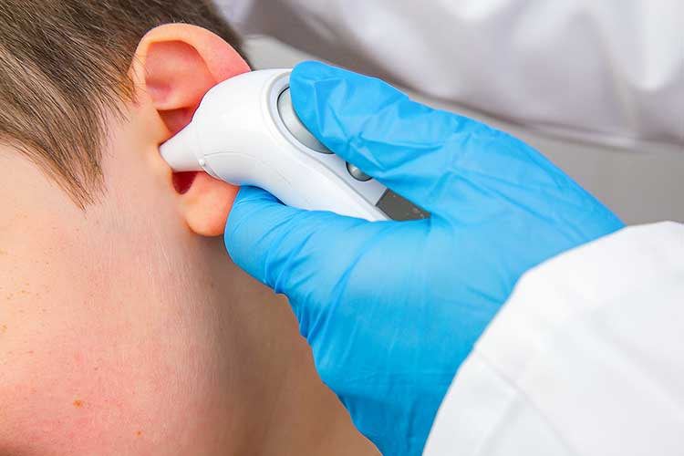
Tympanic Route
- Remove any items that have been covering the patient’s ear (e.g. beanies, headphones) and wait until their ear cools down before proceeding.
- If the patient has been wearing a hearing aid, remove it and wait 20 minutes before taking their temperature.
- Place a protective covering over the probe.
- Ensure the patient keeps their head still.
- Pull the helix up and back (or down and back for babies younger than 12 months) in order to visualize the ear canal.
- Center the probe in the ear and gently insert it inward towards the eardrum. Do not force it in or obstruct the ear canal.
- Keep the probe in place until the thermometer beeps (refer to the manufacturer’s instructions).
- Carefully remove the probe from the ear.
- Record the temperature reading on the display panel.
- Discard the probe covering.
- Ensure the probe is clean and return it to the holder.
(Lapum et al. 2018; Healthwise 2022; RCHM 2019; QAS 2021a)
Forehead Route (Using a Non-contact Infrared Thermometer)
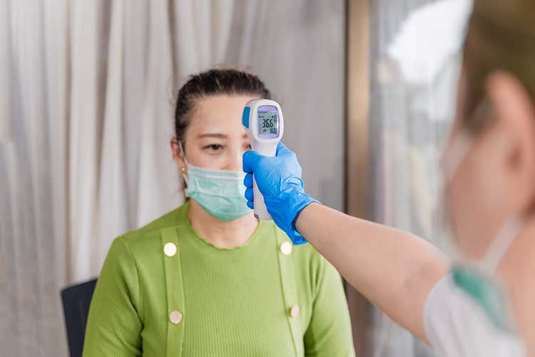
- Ensure you are in a draft-free area, out of direct sunlight. Ideal conditions are between 16ºC and 40ºC with under 85% humidity.
- The device should be left to adjust in the chosen environment for 10 to 30 minutes before use.
- Ensure the patient’s forehead is clean, dry, and unobstructed.
- Check whether the patient has been wearing head covers (e.g. headbands or hats) or facial cleansing products (e.g. cosmetic wipes) that may interfere with the measurement.
- Hold the sensor perpendicular to the patient’s forehead. The patient should remain still.
- Refer to the manufacturer’s instructions for the specific device you are using.
- Do not touch the sensor area. Ensure it stays clean and dry.
(FDA 2023)
Interpreting Thermometer Results
Fever (Pyrexia)
A fever is generally defined as a body temperature of over 38°C (Healthdirect 2020). It is a purposeful increase in body temperature by the hypothalamus, generally in response to infection, to make the body less inhabitable for infectious agents (Roland & Sampson 2020; Swaminathen 2021).
A fever may indicate a range of conditions, including:
- Viral respiratory tract infections
- Ear infections
- Gastroenteritis
- Urinary tract infections
- Pneumonia
- Meningitis
(Healthdirect 2023)
Hyperthermia
Read: Hyperthermia and Heatstroke: Caring for Older Adults in Summer
Unlike a fever, hyperthermia is not a controlled response by the body. Instead, it is a failure of the body’s thermoregulatory systems, resulting in the inability to adequately dispel heat (Swaminathen 2021).
Hyperthermia may be caused by external factors such as excessive environmental heat or a condition such as a pheochromocytoma that causes increased internal heat production (Swaminathen 2021).
Symptoms of hyperthermia may include:
- Headache
- Dizziness
- Sweating
- Nausea and vomiting
- Muscle pain and cramps
- Fatigue
(QAS 2021b)
Severe hyperthermia (known as heatstroke) is defined as a body temperature of over 40°C. This is a medical emergency that may lead to multi-organ failure and death.
The symptoms of heatstroke may be similar to those of less severe hyperthermia. However, a key difference is that a patient with heatstroke may not sweat (Better Health Channel 2022).
It’s important to note that sepsis may be associated with an extremely high fever (potentially up to 40.5°C). If a patient presents with a temperature this high, it’s essential to determine whether they are experiencing sepsis or severe hyperthermia (Swaminathen 2021).
Note that sepsis can present in multiple ways, and body temperature should not be considered the sole or primary indicator for sepsis. It is important to refer to the current sepsis guidelines assessment practices.
For more information, see: Sepsis: Signs and Symptoms.
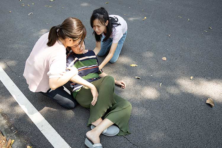
Hypothermia
Hypothermia is defined as a body temperature of under 35ºC. It is most often caused by external factors such as prolonged exposure to cold weather (Mayo Clinic 2022; Lapum et al. 2018).
Other causes include heat loss from lost skin integrity or trauma, decreased heat production due to age and dysfunction of the central nervous system (e.g. stroke) (QAS 2021c).
Hypothermia is a medical emergency that may lead to cardiac and respiratory system failure and, consequently, death (Mayo Clinic 2022). The following symptoms may indicate hypothermia:
- Shivering
- Slurred or mumbled speech
- Slow and shallow breathing
- Weak pulse
- Clumsiness and impaired coordination
- Drowsiness
- Lack of energy
- Confusion or impaired memory
- Loss of consciousness
(Mayo Clinic 2022)
In extremely severe cases, the patient may go into a stupor or become unresponsive, experience apnoea and display very little signs of life (QAS 2021c).
If hypothermia is suspected, preventing further heat loss and rewarming the patient is critical (QAS 2021c).
Conclusion
Despite being a simple and quick procedure, measuring a patient’s temperature can be highly useful in detecting fever, hyperthermia or hypothermia and contextualising their overall state of health.
Note: This article is intended as a refresher and should not replace best-practice care. Always refer to your organisation's policy on measuring and interpreting a patient’s temperature.
Test Your Knowledge
Question 1 of 3
A patient presents with a temperature of over 40°C. This could potentially indicate:
Topics
References
- Better Health Channel 2022, Heat-related Illness, Victoria State Government, viewed 22 November 2023, https://www.betterhealth.vic.gov.au/health/healthyliving/heat-stress-and-heat-related-illness
- Healthdirect 2023, Fever, Australian Government, viewed 22 November 2023, https://www.healthdirect.gov.au/fever
- Healthwise 2022a, Body Temperature, University of Michigan Health, viewed 22 November 2023, https://www.uofmhealth.org/health-library/hw198785
- Healthwise 2022b, Fever Temperatures: Accuracy and Comparison, Healthwise, viewed 22 November 2023, https://www.mottchildren.org/health-library/tw9223
- Lapum, JL, Verkuyl, M, Garcia, W, St-Amant, O & Tan, A 2018, Vital Sign Measurement Across the Lifespan – 1st Canadian Edition, eCampusOntario, viewed 22 November 2023, https://opentextbc.ca/vitalsign/
- Mayo Clinic 2022, Hypothermia, Mayo Clinic, viewed 22 November 2023, https://www.mayoclinic.org/diseases-conditions/hypothermia/symptoms-causes/syc-20352682
- Mayo Clinic 2023, Thermometers: Understand the Options, Mayo Clinic, viewed 22 November 2023, https://www.mayoclinic.org/diseases-conditions/fever/in-depth/thermometers/art-20046737
- Physiopedia 2022, Vital Signs, Physiopedia, viewed 22 November 2023, https://www.physio-pedia.com/Vital_Signs
- Queensland Ambulance Service 2021b, Clinical Practice Guidelines: Environmental/Hyperthermia, Queensland Government, viewed 22 November 2023, https://www.ambulance.qld.gov.au/docs/clinical/cpg/CPG_Hyperthermia.pdf
- Queensland Ambulance Service 2021c, Clinical Practice Guidelines: Environmental/Hypothermia, Queensland Government, viewed 22 November 2023, https://www.ambulance.qld.gov.au/docs/clinical/cpg/CPG_Hypothermia.pdf
- Queensland Ambulance Service 2021a, Clinical Practice Procedures: Assessment/Temperature - Braun ThermoScan® Pro 4000, Queensland Government, viewed 22 November 2023, https://www.ambulance.qld.gov.au/docs/clinical/cpp/CPP_Temperature_Braun%20ThermoScan%20Pro%204000.pdf
- RNpedia 2017, Assessing Body Temperature, RNpedia, viewed 22 November 2023, https://www.rnpedia.com/nursing-notes/fundamentals-in-nursing-notes/assessing-body-temperature/
- Roland, J & Sampson, S 2020, ‘What Is Hyperthermia and How Is It Treated?’, Healthline, 21 August, viewed 22 November 2023, https://www.healthline.com/health/hyperthermia
- Royal Children’s Hospital Melbourne 2019, Temperature Management, RCHM, viewed 22 November 2023, https://www.rch.org.au/rchcpg/hospital_clinical_guideline_index/Temperature_management/
- SafeWork New South Wales 2020, Core Body Temperature v Peripheral Body Temperature, New South Wales Government, viewed 22 November 2023, https://www.safework.nsw.gov.au/resource-library/heat-and-environment/working-in-extreme-heat-the-facts/accordians/core-body-temperature
- Swaminathen, A 2021, ‘Pyrexia, Fever, Hyperthermia - What is the Difference?’, ONiO, 18 February, viewed 22 November 2023, https://www.onio.com/article/pyrexia-fever-hyperthermia-what-is-the-difference.html
- U.S. Food and Drug Administration 2023, Non-contact Infrared Thermometers, FDA, United States Government, viewed 22 November 2023, https://www.fda.gov/medical-devices/general-hospital-devices-and-supplies/non-contact-infrared-thermometers
- Vandergriendt, C, Wright, SA & Stephens, C 2022, ‘What Is the Normal Body Temperature Range?’, Healthline, 5 January, viewed 22 November 2023, https://www.healthline.com/health/what-is-normal-body-temperature